Figure 6.
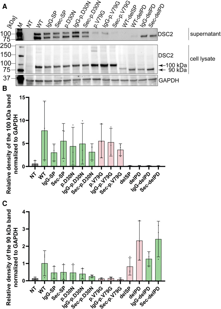
Western blot analysis of secretion assay. (A) Cell culture supernatant on top and HEK293 c18 lysate (bottom). Primary antibody was used against DSC2. As loading control for the cell lysate GAPDH was used. Bar graphs showing relative density of DSC2 constructs normalized to GAPDH (B) at 100 kDa and (C) at 90 kDa. Green bar graphs represent variants which were efficiently expressed and red bar graphs show variants which were expressed but not efficiently secreted. delPD, deleted prodomain of DSC2; delSP, deleted signal peptide of DSC2; M, Marker, NT, non-transfected (negative control); WT, wildtype DSC2.

