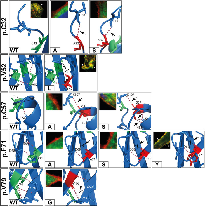
Figure 7.
In silico predictions of polar contacts (red dashed line, arrow) in wildtype DSC2 compared to different prodomain variants. Variants that are not shown do not show different polar contacts in silico (see also Table 2). Conserved amino acids are shown in green and variants in red. Interacting amino acids are shown as sticks and are labeled. Intramolecular bonds are marked with a black arrow. The loss of a polar contact is marked with “X”. Fluorescence microscopic representative images show DSC2 prodomain variants (green) and Wheat germ agglutinin (WGA) conjugated with Alexa Fluor 633 as plasma membrane marker (red) in HT-1080 cells. Colocalization of DSC2 prodomain variants and WGA are indicated by white arrows. The variants p.V52l, p.C57A/S and p.F71Y show different intradomain contact sites but were correctly integrated in the plasma membrane. Five variants (p.C32A/S, p.F71A/S, p.V79G) were not integrated in the plasma membrane. One of the variants (p.V79G) is listed in the Human Gene Mutation Database (HGMD) and was not localized at the plasma membrane. In summary, changes of the molecular modeling structures do not correlate with the plasma membrane transport in the investiaged DSC2 constructs.

