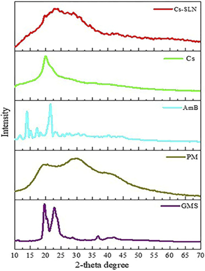
FIGURE 5.
XRD spectra of glycerol monostearate (GMS), amphotericin (AmB), paromomycin, chitosan (Cs), and grafted chitosan-functionalized solid lipid nanoparticles (SLN) of drug (Cs-SLN). X-ray diffractogram of AmB exhibited a sharp peak at the 2θ scattered angle of 21.39, 14.1, and 21.78°, indicating its crystallinity. However, PM was found to be amorphous in nature. Cs-SLN showed two peaks at 24.8° and 30.6°. Nonetheless, the pattern was shifted and broadened, and a weaker peak than GMS was partially recrystallized and transformed to less order in SLN formulation. GMS peaks were also absent due to the complete entrapment of the drug in the lipid matrix. The broadening of the diffraction peaks was attributed to particle sizes as the broadening of Bragg’s peaks indicates the formation of nanoparticles. Adapted with permission from Parvez et al. (2020) under CC BY version 4.0.

