
Figure 1.
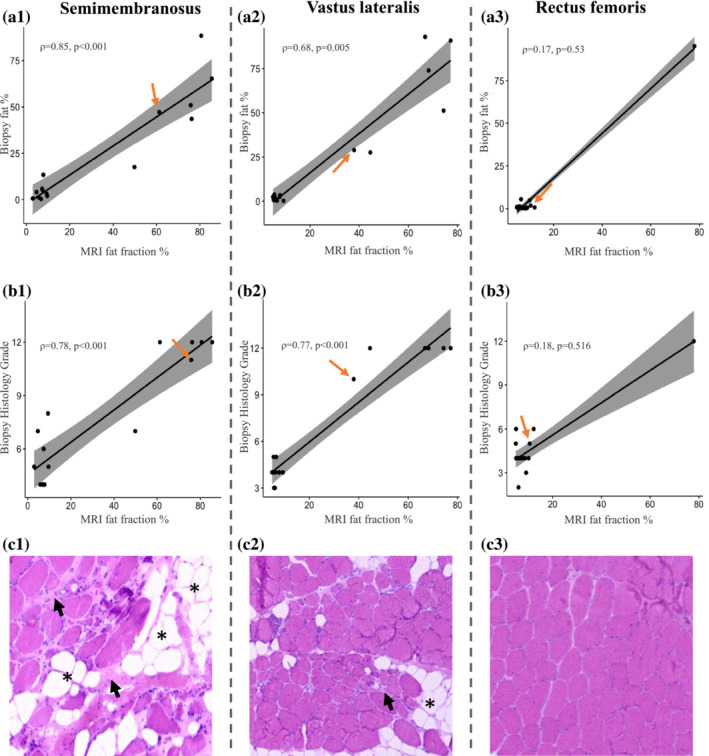
Correlation of PDFF on MRI with muscle biopsy fat percentage and histopathology grading scale. (A, B) There is a strong correlation between the fat fraction measured on MRI and both the fat percentage and severity of histopathological changes on biopsy of the corresponding area of the semimembranosus and vastus lateralis muscles, but not the rectus femoris. The orange arrows indicate the datapoints of the patient whose muscle biopsies are shown in row c. (C) Haematoxylin and eosin‐stained muscle biopsy transverse sections of the respective muscles demonstrate the degree of histopathological changes within one patient: The semimembranosus is the most affected muscle (c1), the vastus lateralis is intermediately affected (c2) and the rectus femoris only mildly affected (c3). The biopsy images show increased fibre size variability, fatty tissue infiltration indicated by the asterisks, and fibrosis shown by the short black arrows. The biopsy images shown are purely for illustrative purposes and represent only a small portion of the analysed muscle tissue.