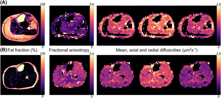
Figure 2.

Representative lower leg ‘Dixon’ chemical‐shift‐based water‐fat separation images and stimulated‐echo diffusion‐tensor magnetic resonance imaging (STE‐DT‐MRI) parameter maps from a 59‐year‐old Becker muscular dystrophy (BMD) patient (A) and a 58‐year‐old male healthy control (B). STE‐DT‐MRI data were acquired with a diffusion time of 330 ms. The BMD patient shows severe fat replacement in the gastrocnemius medialis and lateralis and in the peroneus longus and extensor digitorum longus muscles. This leads to signal voids in these regions in the DT‐MRI data, which were acquired with comprehensive fat suppression. 20
