
Figure 8.
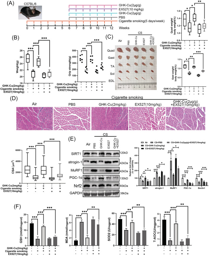
GHK‐Cu protects mice against CS‐induced skeletal muscle dysfunction via SIRT1. (A) Schematic diagram of the control group (exposed to room air), and other four groups of mice exposure to cigarette smoking (5 days/week, 12 weeks) with PBS (every week, 7 weeks; n = 8), high dosage of GHK‐Cu (2 mg/kg body weight every week, 7 weeks; n = 8), EX527 solution (10 mg/kg body weight every week, 7 weeks; n = 8), and high dosage of GHK‐Cu with EX527 solution. (B) The difference of body weight (left) and grip strength (right) between each group. (C) The representative samples of dissected skeletal muscle (left), including Quad (quadriceps), Gast (gastrocnemius), soleus, TA (tibialis anterior), sol (soleus), and EDL (extensor digitorum longus), and the difference of the ratios of Gast and Quad muscle weight to body weight (right). (D) The representative H&E staining of myofibre cross‐section of Gast (upper), and the difference of muscle cross‐sectional area of Gast muscle fibre between each group (lower). (E) Western blot analysis showed the expression of atrogin‐1, MuRF1, PGC‐1α, and Nrf2 levels in mouse Gast muscle; (F) The levels of GSH, MDA, SOD2, and T‐AOC in mouse Gast muscle between each group; *P < 0.05; **P < 0.01; ***P < 0.001.