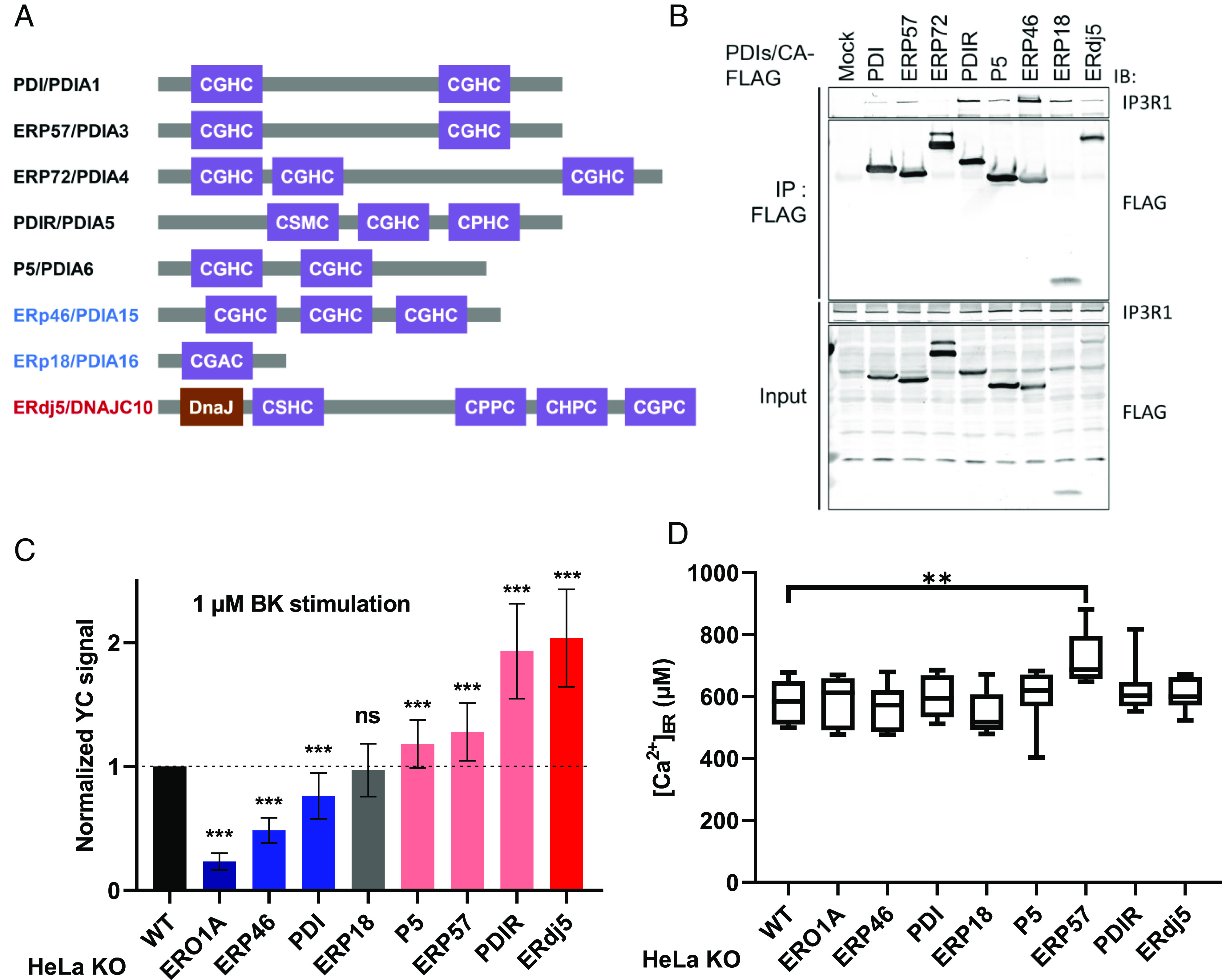
Fig. 1.

The modulation of IICR through PDI family proteins. (A) PDI family proteins (PDIs) examined in this work. The redox-active thioredoxin motif (CXXC) and DnaJ domain are shown. Black: oxidase, reductase, and isomerase. Blue: oxidase. Red: reductase. (B) The interaction of PDIs with IP3Rs. Immunoprecipitates were prepared from HEK-293T cells transiently expressing the indicated FLAG-tagged PDIs and detected by immunoblotting with the indicated antibodies. (C) Quantification of BK-induced IICR in HeLa WT cells or PDI-deficient cells. After transfection with YC3.6, the cells were stimulated with 1 μM BK, and the signal was normalized to the measured peak amplitude. The data present the means ± SD. See SI Appendix, Fig. S1 A and B for the results validating the PDI-deficient cells. (D) The absolute [Ca2+]ER in each KO cell line was calculated from measures of GEM-CEPIA1er ratio. The steady-state [Ca2+]ER in each KO line was determined by Ca2+ titration after sequential treatment with egtazic acid (EGTA) and CaCl2. The horizontal line within the box represents the median value, the upper and lower edges of the box represent 75 and 25% values, and the whiskers represent the total range. ns, not significant; **P < 0.01.
