Fig. 1.

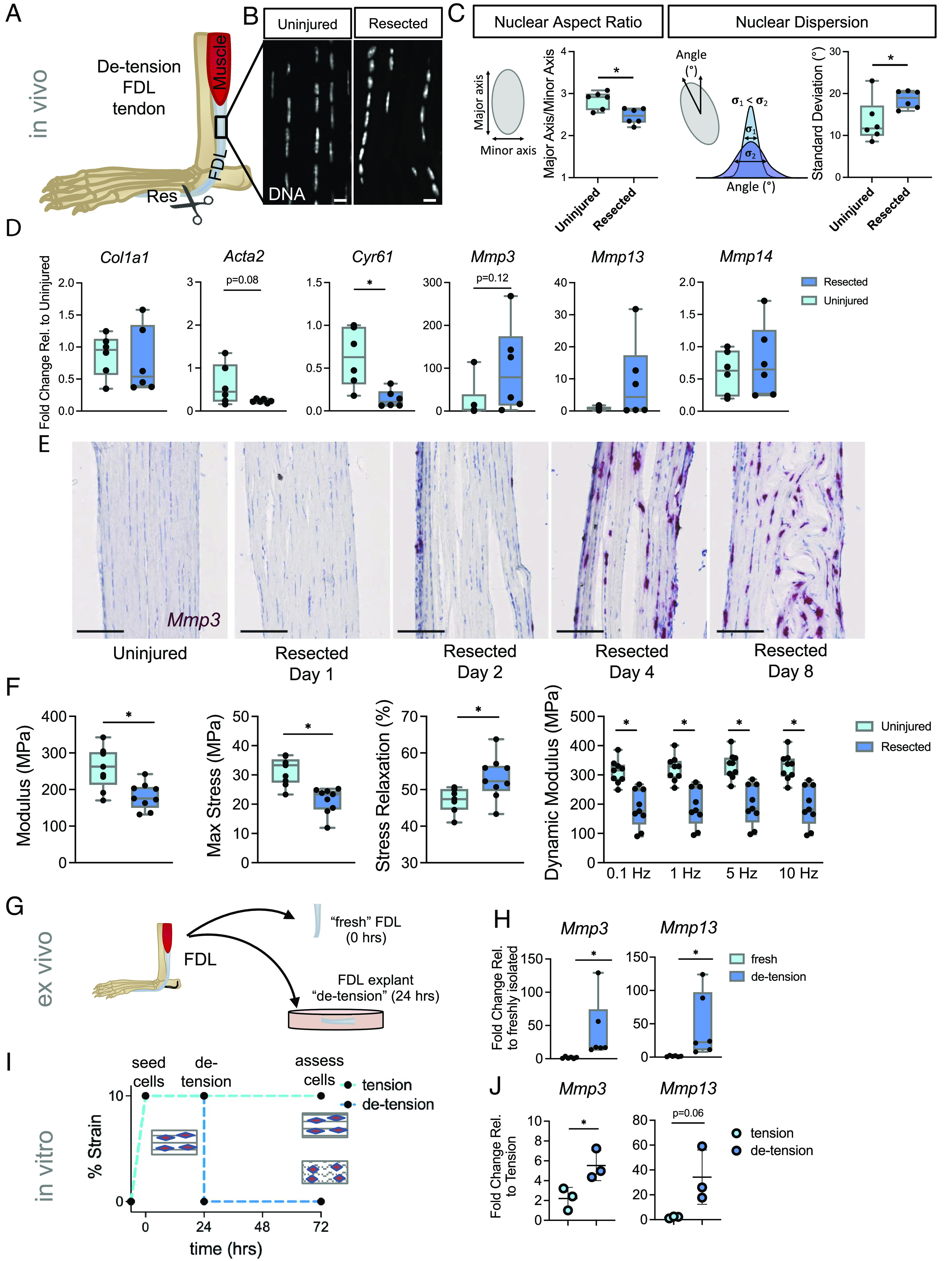
Pathologic acute mechanoresponses of tendon fibroblasts to de-tensioning. (A) Schematic showing approach to de-tensioning the FDL tendon in vivo. (B) DAPI staining depicting nuclear morphology following loss of tension (Scale bar, 10 μm.) (C) Schematic and quantification of nuclear aspect ratio and nuclear dispersion angle of tendon fibroblasts 24 h following loss of tension in the FDL (n = 6 biological replicates). (D) qRT-PCR analysis of FDL tendons 24 h following resection (n = 6 biological replicates). (E) RNAscope analysis for Mmp3 over time following resection of the FDL tendon (Scale bar, 100 μm.) (F) Mechanical properties of resected FDL tendons and their contralateral uninjured controls on day 8 post-resection (n = 9). (G) Schematic showing FDL explant de-tensioning experiment. (H) qRT-PCR analysis of FDL explants (n = 6/group). (I) Schematic showing scaffold de-tensioning experiment (using aligned electrospun scaffolds). (J) qRT-PCR analysis of cells from scaffolds that were either left in tension or de-tensioned for 48 h (n = 3/group). *P < 0.05 evaluated by paired t test. Error bars represent SD.
