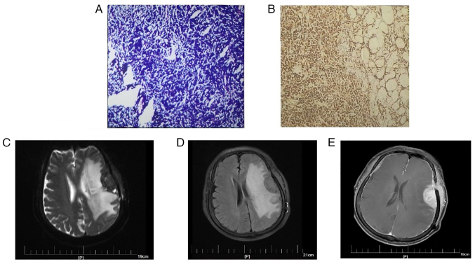
Figure 3.
Pathological findings of tumors and the MRI scans of tumor recurrence. (A) H&E staining of the tumor revealed that the tumor was composed of uniform-sized lymphoid cells with atypia. The tumor cells showed diffuse or flaky growth, with light staining of the cytoplasm, small to medium irregular nuclei, slightly sparse chromatin, and inconspicuous nucleoli. (B) Immunohistochemical findings for CD20: The tumors cells highly expressed CD20. (C) T2 phase, (D) T2FLAIR and (E) enhanced MRI scans. Axial MRI showed abnormal enhancement of left frontotemporal lobe with surrounding edema and multiple enhancement of intracranial meninges as well as left frontotemporal subcutaneous and left buccal lesions.

