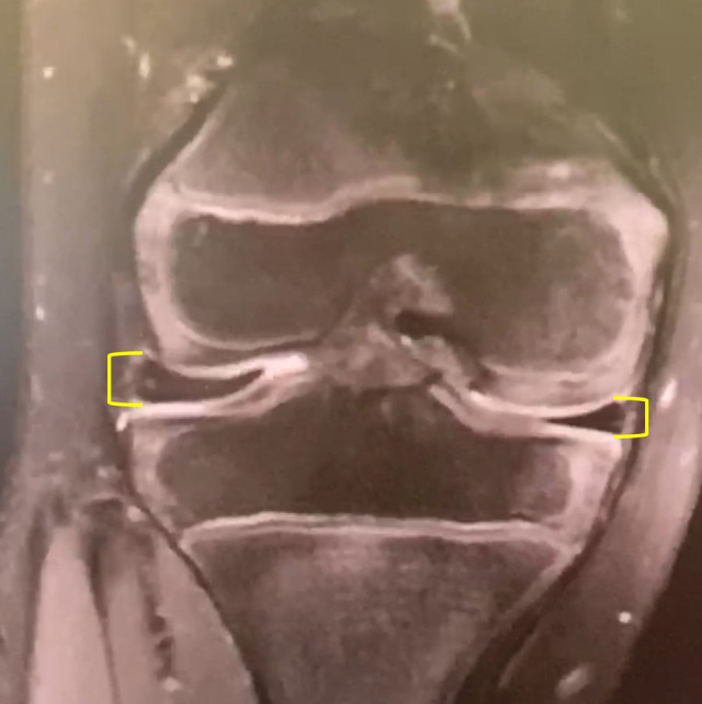
Figure 3.

Discoid lateral meniscus height classification. Coronal cuts were used to compare the discoid lateral meniscus height to the corresponding medial meniscus. Increased height along any portion of the lateral meniscus was classified as abnormal. Yellow brackets indicate the height of the lateral and medial meniscus, demonstrating increased height of the lateral meniscus as compared with the medial meniscus at the peripheral portion of the meniscus.
