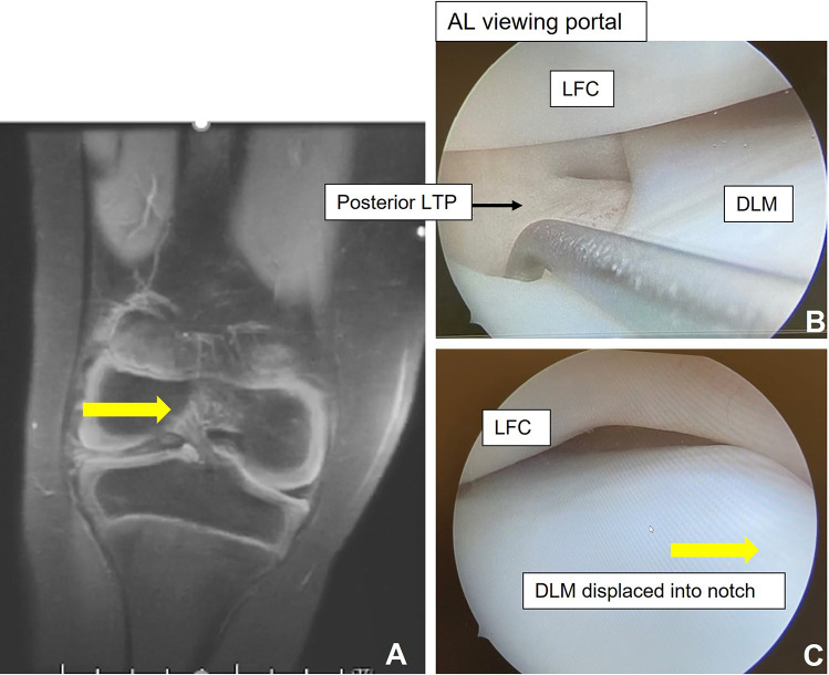
Figure 6.
(A) Coronal magnetic resonance imaging of the right knee demonstrates displacement of the DLM into the notch, with yellow arrow indicating direction of displacement. (B) Arthroscopic comparison of the same knee shows instability along the body and (C) displacement of the DLM into the notch (yellow arrow identifies direction of displacement). AL, anterolateral; LFC, lateral femoral condyle; LTP, lateral tibial plateau.

