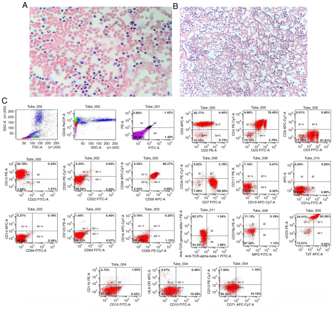
Figure 3.
Papanicolaou staining and flow cytometry results for the present case. Papanicolaou staining revealed no heterotypic cells in the (A) pericardial drainage fluid (magnification, ×200) and (B) thoracic drainage fluid (magnification, ×100). (C) Gate analysis on the CD45/SSC dot plot showed an abnormal cell population in the original cell distribution region, comprising ~58% of the nuclear cells, with positive expression of CD3, CD4, CD5, CD7, CD10, CD38, CD58, TCR γ/δ, cCD3 and TdT. Myeloid proliferation was significantly inhibited. SSC, side scatter; TCR, T-cell receptor; cCD3, cytoplasmic CD3; TdT, terminal deoxynucleotidyl transferase.

