Fig. 1.
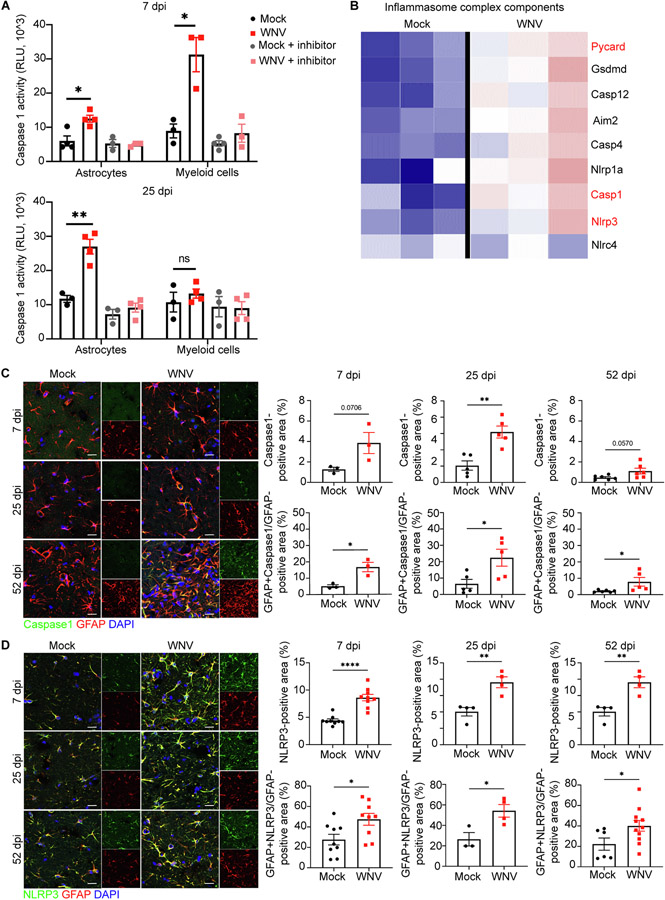
NLRP3/Caspase-1 inflammasome complex is involved in IL-1β production by astrocytes. A. Caspase-1 activity assessment in isolated hippocampal astrocytes and microglia at 7 and 25 dpi using Caspase-1® assay; n = 3–4 animals per group. B. Heat map showing relative expression of significantly altered inflammasome complex genes of mock versus WNV-recovered hippocampi at 25 dpi, each column represents an individual mouse. Font red signify most well-studied inflammasome components. C. Representative immunostaining of Caspase-1 (green), GFAP (red) and DAPI (blue) in hippocampal CA3 region of mock or WNV-infected wildtype animals at 7, 25, and 52 dpi, followed by quantification of percent area of Caspase-1 and GFAP + Caspase-1 + area, normalized to the total GFAP + area; n = 3(7), 5 (25, 52). D. Representative immunostaining of NLRP3 (green), GFAP (red) and DAPI (blue) in hippocampal CA3 region of mock or WNV-infected wildtype animals at 7, 25, and 52 dpi, followed by quantification of percent area of NLRP3 and GFAP + NLRP3 + area, normalized to the total GFAP + area; n = 9 (7dpi), 3–4 (25dpi), 4–11 (52dpi). Data were pooled from at least two independent experiments. Scale bars, 15 μm. Data represent the mean ± s.e.m. and were analyzed by two-way ANOVA and corrected for multiple comparisons.

