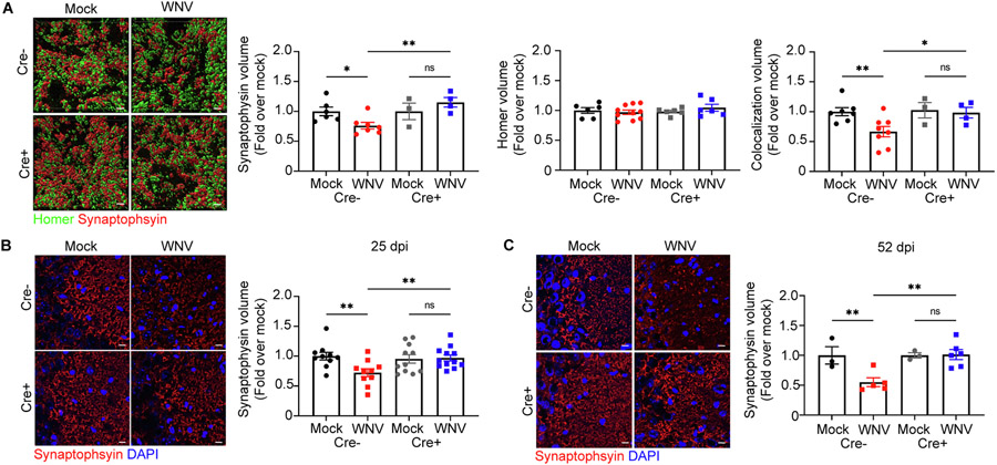
Fig. 4.
WNV-mediated alterations to synapse loss are prevented when IL-1R1 is specifically deleted from NSC. A. Representative 3D rendering of synapses in mock- or WNV-infected Cre− or CreERT + animals at 52 dpi, showing staining for homer (green), synaptophysin (red) and DAPI (blue), followed by quantification of synaptophysin+, homer+, and colocalization of synapse and homer volume in the CA3 of the hippocampus; n = 6–7 (Mock Cre−), 7–10 (WNV Cre−), 3–6 (Mock Cre+), 4–6 (WNV Cre+). B,C. Representative immunostaining of synapses in mock- or WNV-infected Cre− or Cre+ animals at 25 (A) and 52 dpi (B), showing staining for synaptophysin (red) and DAPI (blue), followed by quantification of synaptophysin-positive area in the CA3 of the hippocampus; n = 10 (Mock Cre− 25, WNV Cre− 25), 11 (Mock Cre+ 25), 12 (WNV Cre+ 25), 3 (Mock Cre− 52, WNV Cre−), 5 (WNV Cre− 52), 6 (WNV Cre+ 52). Data were pooled from at least two independent experiments. Scale bars, 2 μm (A) or 10 μm (B,C). Data represent the mean ± s.e.m. and were analyzed by two-way ANOVA and corrected for multiple comparisons.

