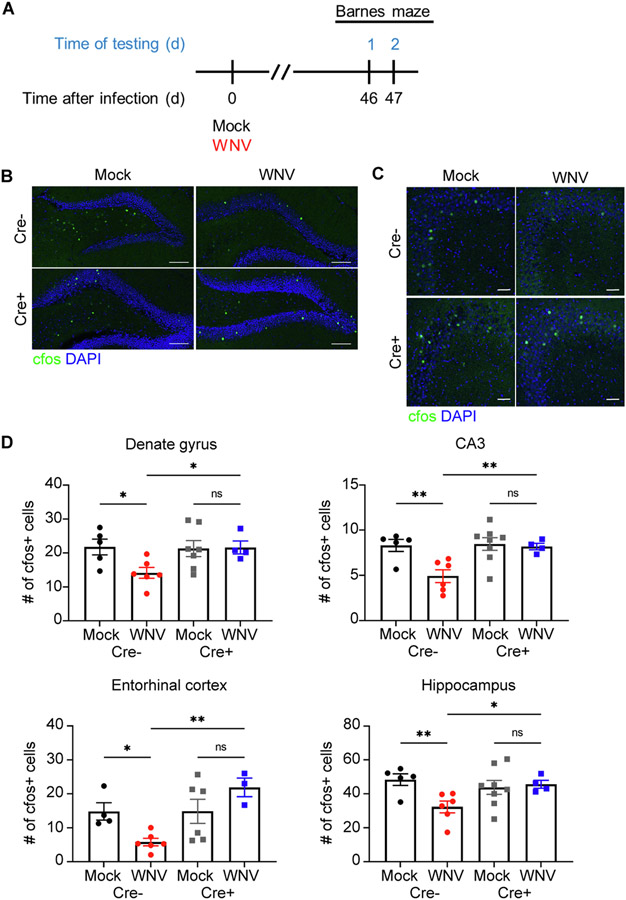
Fig. 6.
Hippocampal neural activity is disrupted in WNV-recovered animals, but loss of IL-1 signaling in NSC protects animals from these alterations. A. Experimental design: mice underwent behavioral testing at 46 dpi, followed by 2 consecutive days of Barnes maze testing. B,C. Microscopy of the dentate gyrus (B) and CA3 (C) region of mock or WNV-infected Cre− or Cre+ animals at 52 dpi, showing staining of CFOS (green) and DAPI (blue). D. Quantification of total CFOS + cells in the DG, CA3 region, entorhinal cortex and the entire hippocampal circuit. Data were pooled from at least two independent experiments; n = 4–5 (Mock Cre−), 5–6 (WNV Cre), 6–8 (Mock Cre+), 3–4 (WNV Cre+). Data represent the mean ± s.e.m. and analyzed by two-way ANOVA and corrected for multiple comparisons.

