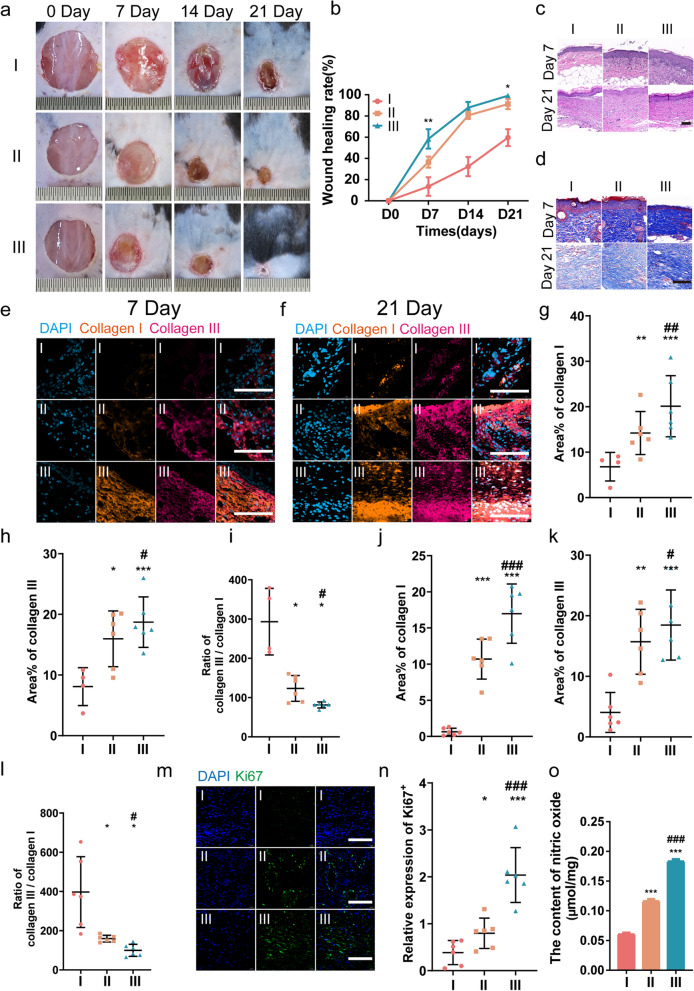
Fig. 5.
UCMSCs-exo/eNOS promote matrix remodeling and tissue repair in diabetic mouse wounds. I: Treatment with PBS. II: Treatment with UCMSCs-exo. III: Treatment with UCMSCs-exo/eNOS. a, b Representative images of full-thickness skin defects and wound-healing rates in diabetic mice receiving multi-point injections of PBS, UCMSCs-exo, and UCMSCs-exo/eNOS at postoperative days 0, 7, 14, and 21 (n = 4 in each group at each time point, two-tailed Student’s t-test). c H&E staining of wound sections treated with PBS, UCMSCs-exo, and UCMSCs-exo/eNOS at postoperative days 7 and 21 days. d Masson staining of wound sections treated with PBS, UCMSCs-exo, and UCMSCs-exo/eNOS at postoperative days 7 and 21. e Immunofluorescence staining of Collagen I and Collagen III in the wounds of the different groups on postoperative day 7. Scale bar: 100 μm. f Immunofluorescence staining of Collagen I and Collagen III in the wounds of different groups on postoperative day 21. Scale bar: 100 μm. g, h, i Immunofluorescence quantification of Collagen I and Collagen III and the Collagen III to Collagen I ratio in e (n = 4 in I group, n = 6 in II group and n = 6 in III group, Mann–Whitney U test). j, k, l Immunofluorescence quantification of Collagen I and Collagen III and the Collagen III to Collagen I ratio in f (n = 6 in each group, two-tailed Student’s t-test). m Immunofluorescence staining of Ki67 in the wounds of the different groups on postoperative day 7. Scale bar: 100 μm. n Immunofluorescence quantification of Ki67 (n = 6 in each group, two-tailed Student’s t-test). o Subcutaneous injection of PBS, UCMSCs-exo, and UCMSCs-exo/eNOS into diabetic mice with chronic wounds and analysis of the nitric oxide content at the wound site (n = 3 in each group, two-tailed Student’s t-test). Data represent means ± SD. *p < 0.05, **p < 0.01, ***p < 0.001 vs. PBS group; #p < 0.05, ##p < 0.01, ###p < 0.001 vs. UCMSCs-exo group

