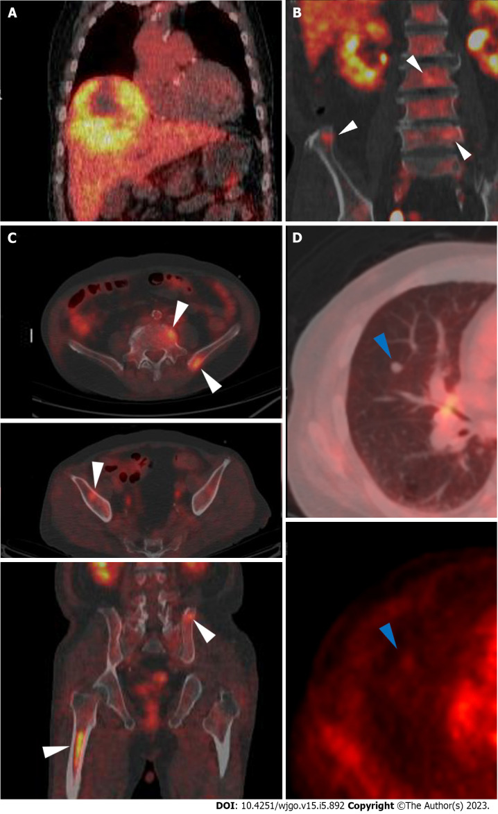
Figure 2.
F-18 fluorodeoxyglucose positron emission tomography/computed tomography images. A: A large hypermetabolic tumor was noted in the right hepatic lobe [maximum standardized uptake value (SUVmax) 5.1]; B and C: Multiple hypermetabolic lesions (SUVmax 4.8) are seen in both iliac bones, lumbar vertebrae, and the right femur (white arrowheads); D: A solid nodule with mild hypermetabolic activity was noted in the right middle lobe (blue arrowheads).

