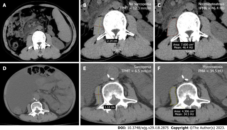
Figure 1.
Abdominal computed tomography images at the level of the third lumbar vertebrae were used to measure the transversal psoas muscle thickness and psoas muscle attenuation in a 45-year-old man without sarcopenia and myosteatosis, and a 51-year-old man with sarcopenia and myosteatosis. A: The axial computed tomography (CT) scan image of a 45-year-old cirrhotic patient (height: 1.74 m, weight: 74 kg) who developed variceal rebleeding; B and C: The patient had normal transversal psoas muscle thickness (TPMT) and psoas muscle attenuation (PMA) levels: 12.3 mm/m (21.4 mm/1.74 m) and 46.4 HU, respectively; D-F: The axial CT scan image of a 51-year-old cirrhotic patient (height: 1.72 m, weight: 55 kg) who developed refractory ascites (D), and had low TPMT and PMA (6.5 mm/m [11.2 mm/1.72 m] and 34.5 HU) (E and F). TPMT: Transversal psoas muscle thickness.

