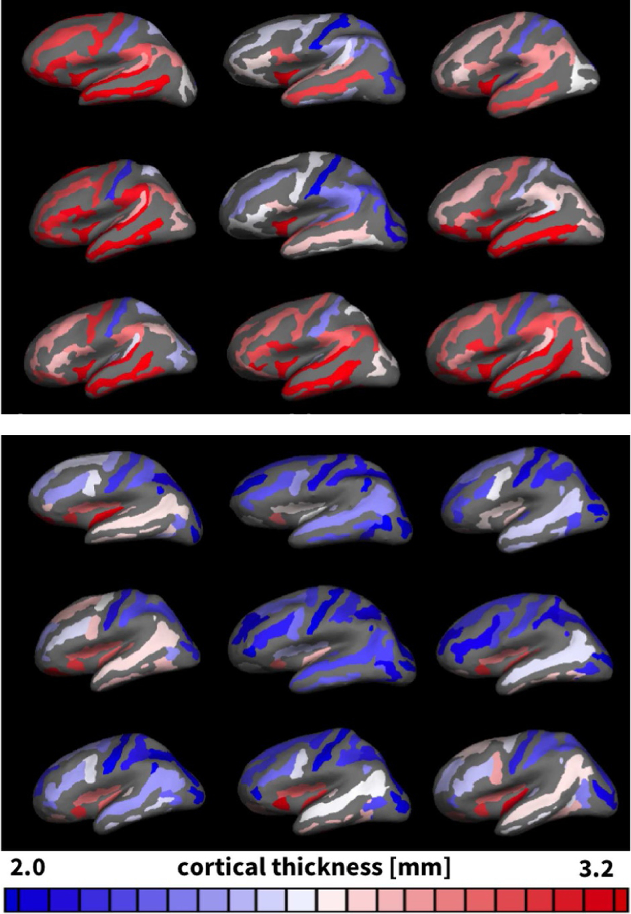
Fig. 4.

Thickness variations in adult human brains. Gyral (top) and sulcal (bottom) regions of n = 9 adult human brains reconstructed, parcellated, and inflated to display the complete pial surface. Regions are color-coded according to the average gyral and sulcal thicknesses in each region; all remaining regions are shown in gray.
