FIGURE 3.

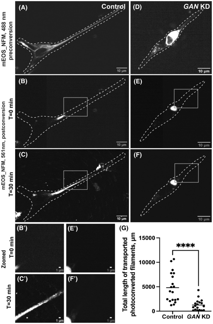
Neurofilament transport is inhibited in GAN KD primary DRG neurons. Photoconverstion of mEOS_NFM in primary DRG neurons using spinning disk confocal microscopy in control (A–C) and GAN KD (D–F) cells. Panel (A) and (D) were imaged under the green channel (488 nm) before photoconversion. Panel (A) shows the NFM filament in a control cell while panel (D) displays the aggregated NFM filament in GAN KD cells. mEOS‐NFM was photoconverted from green to red at the specific region. Panel (B) and (E) were imaged under red channel at time 0 min after photoconversion. Panel (C) and (F) 30 min after photoconversion. Dotted lines mark the boundary of the cell. Gray box regions were zoomed in and shown below (B', C', E' and F'). Scale bar 10 μm for full images and 1 μm for zoomed images. (G) Photoconverted NFM IFs outside the conversion zone were quantified and segmented filaments were counted for each frame. Statistical significance was determined using Student's t‐test (n = 18 cells). ****p < 0.0001.
