Figure 4.

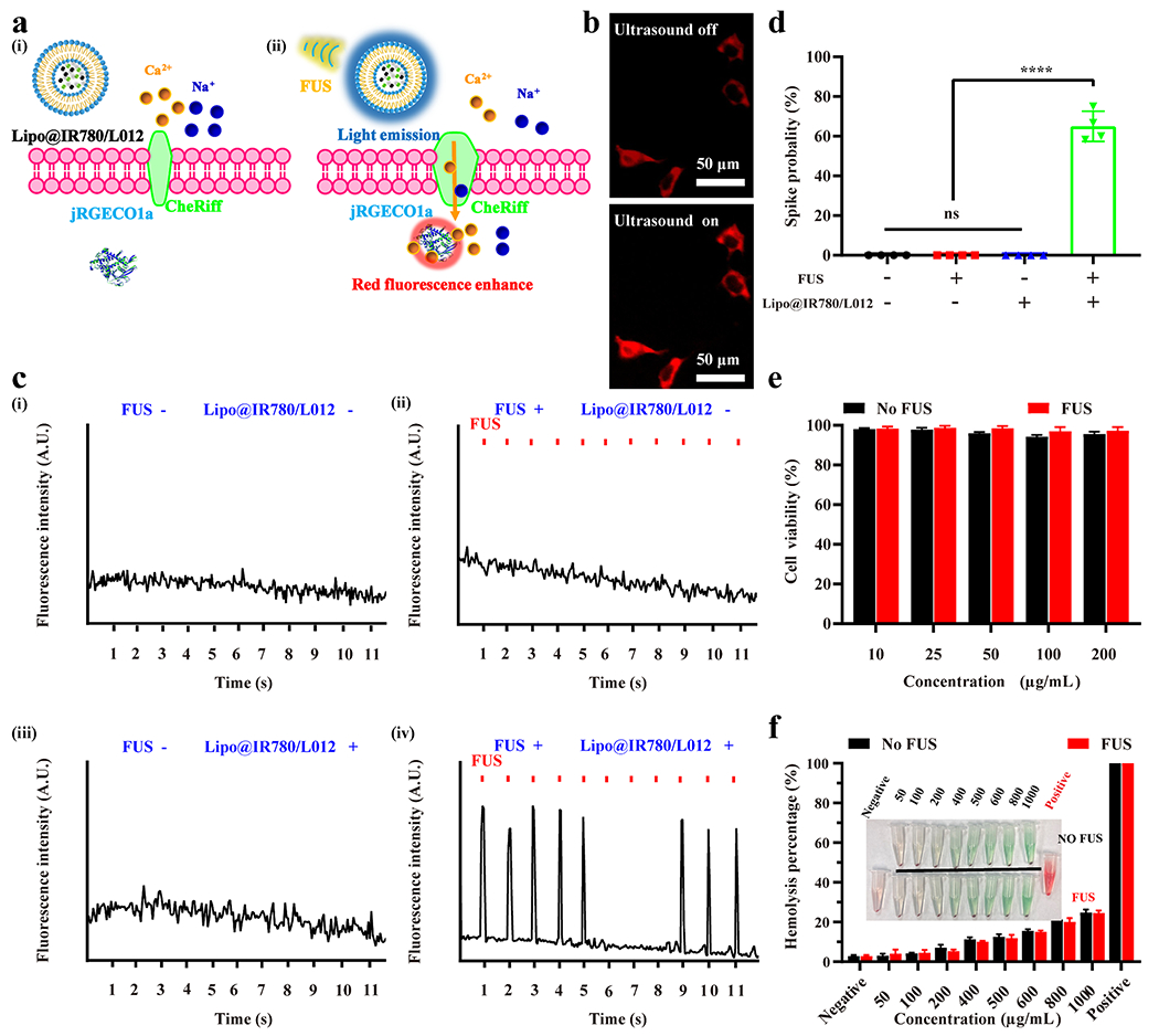
In vitro sono-optogenetic stimulation and biosafety tests of Lipo@IR780/L012. (a) Illustration of FUS-triggered CheRiff channels opening due to 470 nm blue light emission from Lipo@IR780/L012 nanoparticles. The flow of Ca2+ into the cells binds with jREGCO1a proteins to enhance the red fluorescence signal. (b) Fluorescence images of CheRiff-expressing spiking HEK cells with and without mechanoluminescence irradiation from Lipo@IR780/L012 nanoparticles: ultrasound off and ultrasound on; (c) fluorescence signal recording from CheRif-expressing spiking HEK cells under the following conditions: (i) no FUS and no Lipo@IR780/L012 nanoparticles; (ii) with FUS (1.5 MHz, puls 100 ms on, 900 ms off, 1 Hz, 1.5 MPa) and no Lipo@IR780/L012 nanoparticles; (iii) no FUS and with Lipo@IR780/L012 nanoparticles; (iv) with FUS (1.5 MHz, puls 100 ms on, 900 ms off, 1 Hz, 1.5 MPa) and with Lipo@IR780/L012 nanoparticles; and (d) spike probability of CheRiff-expressing spiking HEK cells under the different conditions (n = 4 per group, two-way ANOVA). (e) Cell viability tests of Lipo@IR780/L012 nanoparticles in HEK cells with and without FUS irradiation (n = 5 per group). (f) Hemolysis tests of Lipo@IR780/L012 nanoparticles (n = 3 per group). All plots show mean ± SEM unless otherwise mentioned. *P < 0.05, **P < 0.01, ***P < 0.001, and ****P < 0.0001; ns, not significant.
