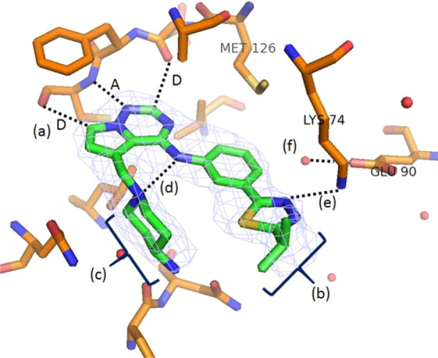
Fig. 2.

X-ray co-crystal structure of 2 bound to AAK1 (PDB ID 8GMC). a Donor-acceptor-donor (D-A-D) interaction with hinge region residues; b hydrophobic interactions with p-loop; c binding of amino piperidine in sugar pocket; d H-bond between the piperidine nitrogen and the anilinic N-H; e H-bonding between thiadiazole nitrogen and Lys74; and f water molecule bound to Glu90. Water molecules are shown as red spheres
