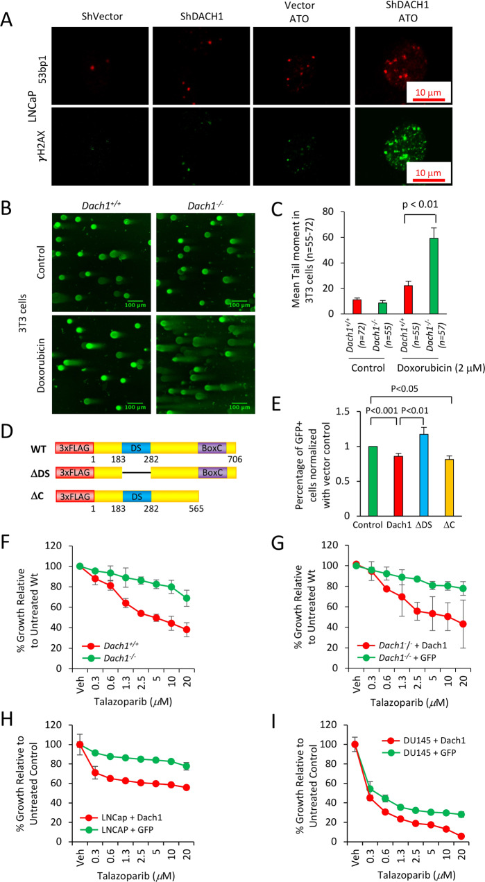
Fig. 7. DACH1 enhances DNA repair.
A LNCaP cells stably transduced with control vector or shDACH1 were treated with ATO (1 μM), and immunofluorescence for 53BP1 or γH2AX was conducted. (B) The neutral pH comet assay, which mainly detects DNA double-strand breaks (DSBs), was conducted as a single-cell DNA damage assay. Dach1+/+ and Dach1−/− 3T3 cells were treated with 2 μM doxorubicin for 18 h. Scale bar, 100 μm with (C) data shown as mean ± SEM. D Schematic representation of DACH1 expression vectors, which were introduced into (E) U2OS cells expressing I-SceI based reporter assays for homologous repair (DR-GFP). Cells were analyzed after 48 h with data shown as mean ± SEM for N = 12 (DACH1), N = 7 (DACH1 ΔDS), and N = 5 (DACH1ΔC). F Dach1+/+ and Dach1−/− 3T3 cells or (G) Dach1−/− 3T3 transduced with a MSCV/DACH1-IRES-GFP expression vector or GFP vector control, were treated for 3 days with increasing doses of Talazoparib, a PARP inhibitor. Data are shown as mean ± SEM for triplicate (N = 3). H, I LNCaP (H) and DU145 (I) cells were transduced with a pLRT-DACH1 expression vector or vector control. After DACH1 induction with doxycycline (1 mg/mL), cells were treated for 3 days with increasing doses of Talazoparib. Cell number was determined by methylene blue assay using a standard curve for each cell line as a reference. Data are shown as mean ± SEM for triplicates (N = 3).

