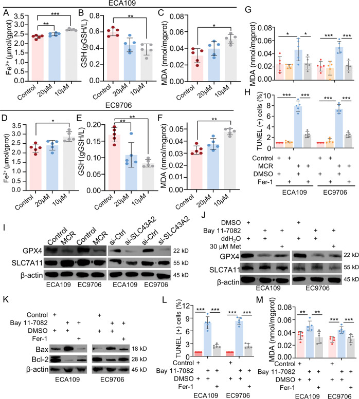
Fig. 6. MCR-induced ferroptosis by inhibited GPX4 expression in ESCC.
A–F The expressions of ferroptosis-related indexes in ECA109 and EC9706 cell lines were detected in control group, 10 μM group and 20 μM group. For 10 μM group or 20 μM group, 10 μM or 20 μM methionine was added into methionine/cystine-deficient medium, respectively. Cells were cultured for 72 h, and the content of Fe2+ (A, D), GSH (B, E), and MDA (C, F) in cell lysates was detected by ELISA. n = 5 for each group, *P < 0.05, **P < 0.01, ***P < 0.001, two-tailed unpaired Student’s t test. G, H The content of MDA (G) and the TUNEL-positive cells (H) were detected respectively in (1) control+DMSO group; (2) control+Fer-1 group; (3) MCR + DMSO group; (4) MCR+Fer-1 group. n = 5 for each group, *P < 0.05, ***P < 0.001, two-tailed unpaired Student’s t test. I The expression of GPX4 and SLC7A11 were detected in ESCC cells under MCR condition and transfected with SLC43A2 siRNA by western blot. J Western blot was used to detect the GPX4 and SLC7A11 expression in (1) DMSO+ddH2O group; (2) Bay 11-7082+ddH2O group; (3) Bay 11-7082 + 30 μM group. K The protein level of Bax and Bcl-2 were detected respectively in (1) control+DMSO group; (2) Bay 11-7082 + DMSO group; (3) Bay 11-7082+Fer-1 group. L, M The TUNEL-positive cells (L) and the content of MDA (M) were detected, respectively, in (1) control+DMSO group; (2) Bay 11-7082 + DMSO group; (3) Bay 11-7082+Fer-1 group. n = 5 for each group, *P < 0.05, ***P < 0.001, two-tailed unpaired Student’s t test. Data are presented as mean ± SD (n = 5).

