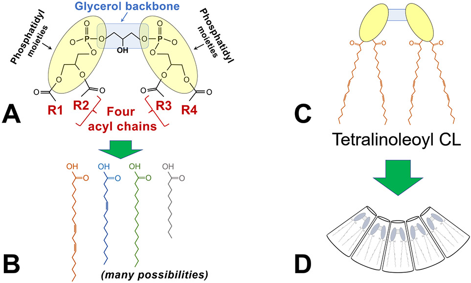
FIGURE 1. Cardiolipin structure, an overview.
A) The schematic shows the key features of cardiolipin: A glycerol backbone and two phosphatidyl moieties that together form the dimeric phosphatidylglycerol head group; and 4 acyl chains (designated R1-R4).
B) Each of the four acyl chains may vary by length, degrees of saturation, and positions of double bonds, resulting in an enormous possible diversity of acyl chain combinations.
C) The most abundant form of cardiolipin in the mammalian heart is tetralinoleoyl cardiolipin (CL), in which the four acyl chains are linoleic acid.
D) Tetralinoleoyl CL has a unique conical shape that is critical to its biophysical properties within the lipid membrane.

