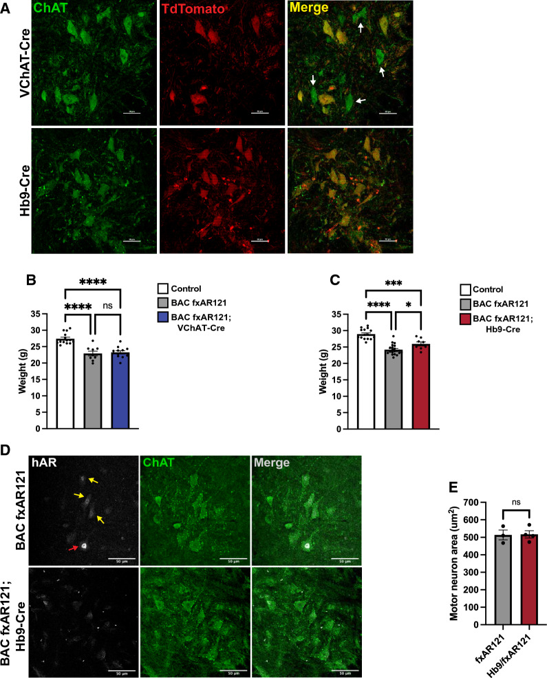
Fig. 4.
Motor neuron-rescued SBMA mice do not display improvements in disease phenotypes. A Representative images of spinal cord sections from VChAT-Cre; TdTomato and Hb9-Cre; TdTomato mice stained for acetylcholine transferase (ChAT), a marker of mature motor neurons. White arrows denote ChAT + motor neurons lacking TdTomato and thus Cre activity. Scale bar = 50 µm. B Weight of 26-week-old male BAC fxAR121; VChAT-Cre mice compared to singly transgenic BAC fxAR121 and control littermates, n = 9–14 mice/group. One-way ANOVA with post-hoc Tukey test, ****P < 0.0001. C) Weight of 26-week-old male BAC fxAR121; Hb9-Cre mice compared to singly transgenic and control littermates, n = 8–21 mice/group. One-way ANOVA with post-hoc Tukey test, *P < 0.05, ***P < 0.001, ****P < 0.0001. D) Representative images of spinal cord sections from symptomatic 9–10 month-old BAC fxAR121 mice and unaffected BAC fxAR121; Hb9-Cre mice stained for mutant human AR (white) and ChAT (green). White arrow denotes motor neurons with strong polyQ-hAR expression, while yellow arrows denote motor neurons with moderate expression. Scale bar = 50 µm. E) Quantification of motor neuron area in lumbar spinal cord of symptomatic 9–10 month-old BAC fxAR121 mice and unaffected BAC fxAR121; Hb9-Cre mice; n = 68–100 motor neurons/animal, 3–4 mice/group. Nested t-test, P = n.s. Error bars = s.e.m

