
Figure 3. Spectral karyotype and HPV in situ hybridization of SNU-1000 and SNU-1245.
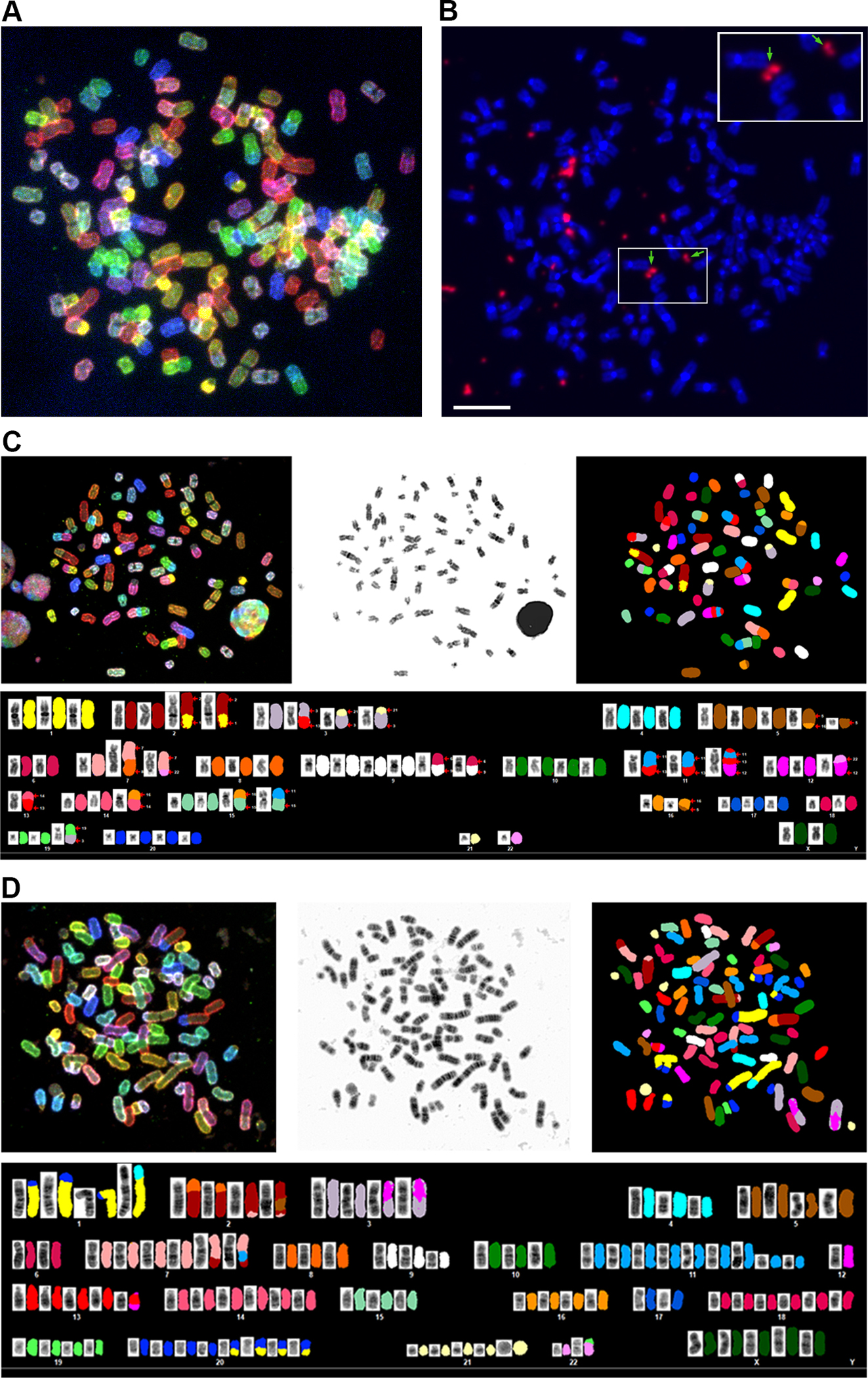
A. A representative spectral karyotype of SNU-1000 cell is shown revealing a near-triploid chromosome content. B. Fluorescent in situ hybridization (FISH) of the same metaphase with a pan-HPV probe reveals intense signals for HPV (in orange) in extrachromosomal regions between or adjacent to chromosomes. Arrows and the inset show HPV signals resembling double minute-like structures. The scale bar is 10 um. C. SKY of SNU-1000 displaying translocation of several chromosomes, including chromosome 11 in the region of the YAP1 amplification. D, SKY of SNU-1245, a near-tetraploid cell line with. Details of metaphases and additional images are in Supplementary Figures S3, S4 and Supplemental Tables 6–8).