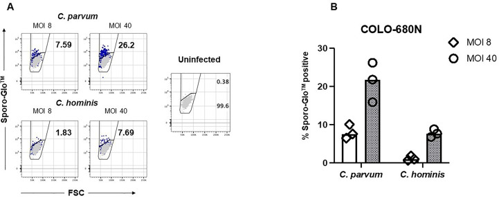
Figure 2.
Sporo-Glo™-positive COLO-680N cells are detectable in increasing proportions with increasing multiplicity of infection (MOI) for C. parvum and C. hominis. (A) COLO-680N cells were infected at the indicated MOI (dark blue dots) with either C. parvum (top two panels) or C. hominis (bottom two panels) and then fixed, permeabilized, and stained with Sporo-Glo™. Uninfected cells stained with Sporo-Glo™ (light gray dots) are overlaid on each panel. A region defining Sporo-Glo™-positive cells is shown, and the percentage of cells in each region is displayed. (B) Triplicates from independent cultures from the experiment shown in (A) plotted as percent Sporo-Glo™ cells.

