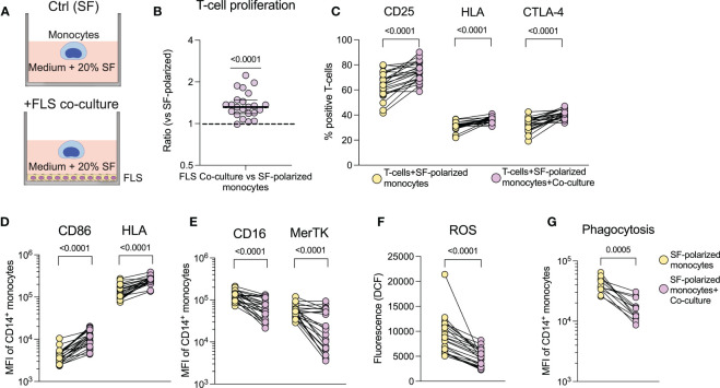
Figure 5.
Increased co-stimulatory capabilities of synovial monocytes are induced in vitro in healthy monocytes through co-culture with fibroblast-like synoviocytes. (A) Experimental setup of the co-culture assay. (B) Monoculture of monocytes (SF) and monocytes co-cultured with FLS (Co-culture) were detached following overnight culture and seeded with CellTrace Violet (CTV) stained anti-CD3 activated T-cells, which were cultured (1:10 monocytes to T-cells) for 72hrs, followed by analysis of proliferation (displayed as ratio of percent proliferation between FLS co-culture vs. monoculture of monocytes) and (C) expression of activation markers in T-cells (n=23). (D) Shows changes in surface expression of CD86 and HLA and (E), of CD16 and MerTK. (F) Displays ROS production after 1hr incubation following H2DCFDA staining (n=23) and (G) phagocytosis of opsonized FITC labelled beads (n=12). Statistics were performed using one sample Wilcoxon signed-rank test (with the hypothetical median of 1) or Wilcoxon matched pairs signed rank test. Lines at median with IQR. FLS, Fibroblast,like synoviocytes; ROS, Reactive oxygen species; MFI, Median fluorescence intensity; SF, Synovial fluid.

