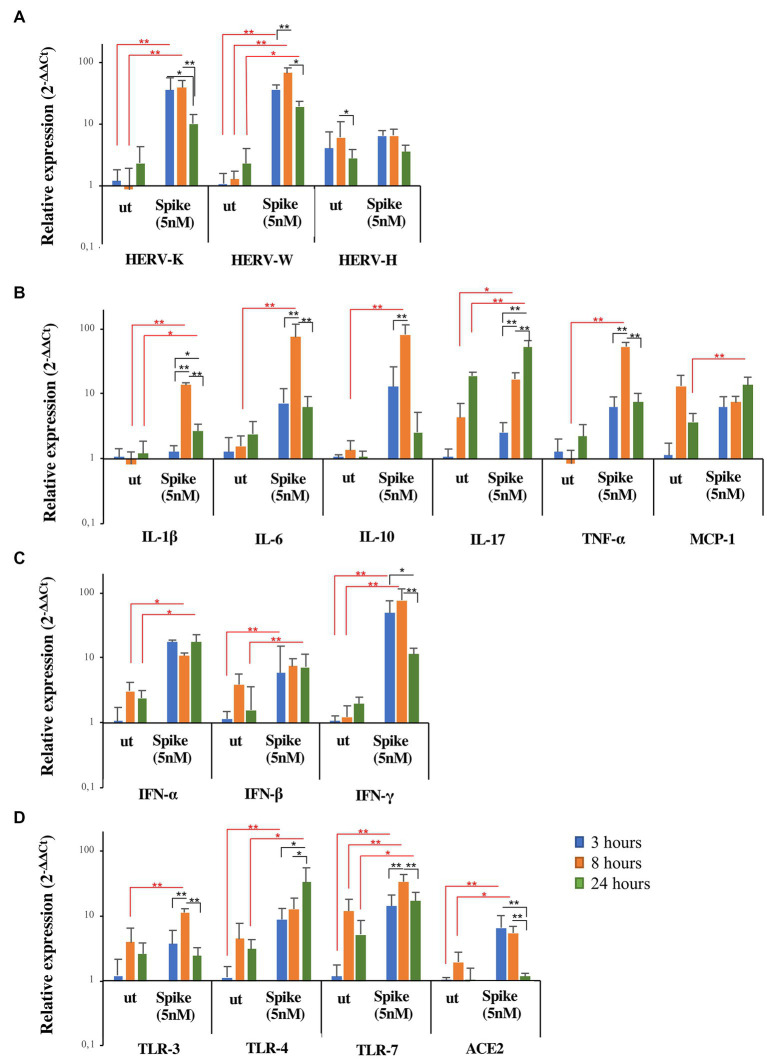
Figure 4.
Expression of HERVs and inflammatory markers in FaDu cells after in vitro stimulation with SARS-CoV-2 spike protein. FaDu cells were stimulated with SARS-CoV-2 spike protein (5 nM) for 3, 8 and 24 h. HERVs (A), cytokines (B), interferons (C) and receptors (D) mRNA levels, obtained by qRT-PCR analysis, were represented as mean ± standard deviation. For comparisons non parametric Kruskal–Wallis test was utilized. Red lines and asterisks outline differences between untreated (ut) and treated with spike protein, while black lines and asterisks delineate differences between spike treatments at different times of exposure.

