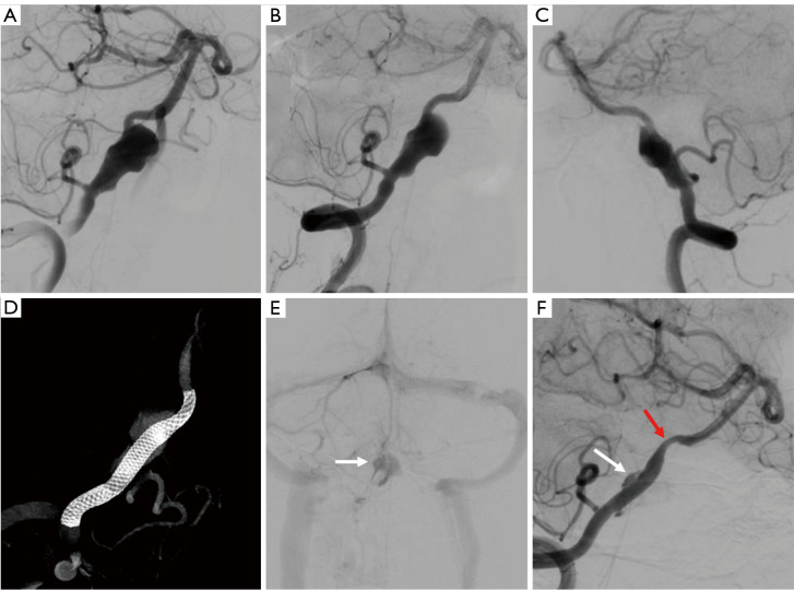
Figure 3.
A typical case in the flow diversion group. (A) Pretreatment DSA showing a right-sided vertebral artery aneurysm. A pipeline flow diverter was deployed without coil embolization, and the immediate postprocedural DSA (B,C) and Vaso CT (D) showed the flow diverter was released properly, without in-stent or distal vessel thrombosis. (E) The postprocedural DSA showed the blood flow was detained to the venous phase. (F) Nine months after the procedure, the DSA showed mild in-stent stenosis and partial contrast filling at the aneurysm neck. The white arrows indicate the aneurysm. The red arrow indicates the in-stent stenosis. DSA, digital subtraction angiography; CT, computer tomography.

