. 2023 Mar 8;48(2):e12. doi: 10.5395/rde.2023.48.e12

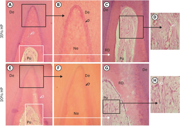
Figure 2. Dentin-pulp complex of mandibular incisors subjected to in-office bleaching treatment (“A-D” = HP35 and “e-h” = HP20). (A) The coronal pulp tissue exhibits a wide area of necrosis (white finger point) associated with notable deposition of tertiary dentin (arrows) (H/E [hematoxylin and eosin], ×40). (B) High magnification of Figure 2A. Note the transition border (black finger point) between the tubular dentin (De) and the necrotic pulp tissue (Ne) (×84). (C) The coronal and root pulp tissue exhibit continuous deposition of reactionary dentin (RD) (H/E, ×125). (D) Detail of the pulp area demonstrated in Figure 2C. The residual pulp tissue shows only a few inflammatory mononuclear cells among dilated and congested blood vessels (white finger point) (H/E, ×250). (E) RD (white finger point) adjacent to the partial coronal pulp necrosis. Note the residual pulp (Pu) tissue (H/E, ×40). (F) High magnification of the tip of pulp horn with necrotic tissue (Ne). Note the transition zone (black finger point) between the tubular dentin (De) and the necrotic tissue (H/E, ×84). (G) Detail of Figure 2E. Subjacent to the tubular dentin (De), a wide layer of RD is observed. Note the residual coronal pulp tissue (Pu) (H/E, ×125. (H) Detail of Figure 2G. Only a few mononuclear inflammatory cells among blood vessels can be seen (H/E, ×250).

Figure 2