
Fig. 2.
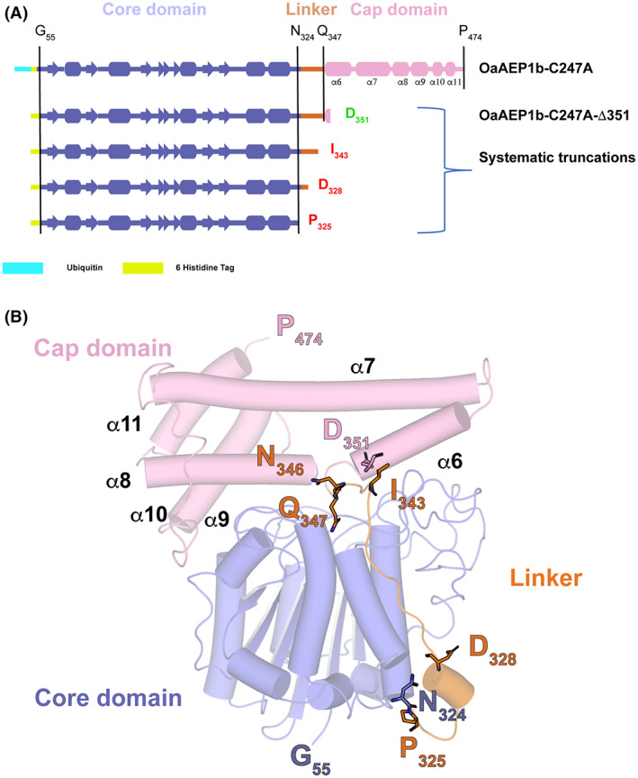
Design of truncation constructs of OaAEP1b (A) Schematic view of the four constructs of OaAEP1b‐C247A that were subjected to expression tests in Escherichia coli in this work. (B) 3D structure of OaAEP1b‐C247A in its proenzyme form (PDB access code: 5H0I) (13). Residues 326–342 from the linker region are flexible and could not be traced in the electron density map. The orange dotted line represents this flexible linker region between the core and cap domains. The truncation sites introduced in our study to obtain a constitutively active peptide ligase are indicated in red.