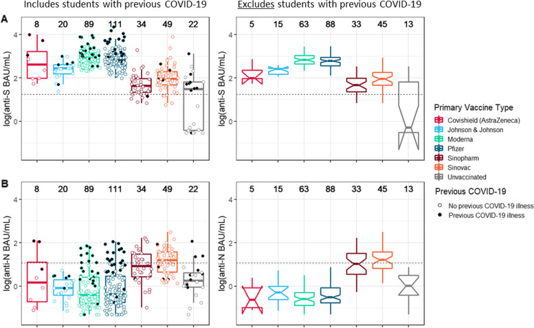
Fig. 1.
Distribution of anti-SARS-CoV-2 IgG antibody levels from sera samples (N = 333) collected from a sample of university students (N = 306) who had only completed a primary series of a COVID-19 vaccine or were unvaccinated — fall academic semester 2021, Wisconsin. Top row, A: antibody levels targeting the spike protein of SARS-CoV-2 (anti-S). Bottom row, B: antibody levels targeting the nucleocapsid of SARS-CoV-2 (anti-N). Within each row, on the left, a scatterplot—shaded according to previous COVID-19 status—and overlaid boxplot displays antibody levels in WHO-standardized binding antibody units per milliliter (BAU/mL), log(10) scale. On the right, notched boxplots are presented, reflecting the 95% confidence intervals around the median value; these only reflect data from students without previous COVID-19. Total datapoints per category are included above each vaccine group. Horizontal dashed lines indicate the assay threshold of positivity. One student (with receipt of Covaxin primary series) not presented

