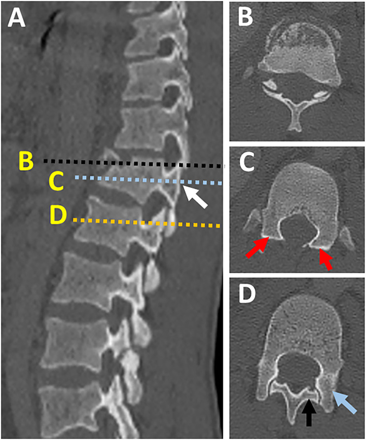
Figure 11.
Naked facet sign. (A) CT parasagittal images showing vertical facet distraction with partial uncoverage of superior articular process (white arrow); (B) Axial CT cut at the level of overlapping facets ( black dashed line in image A) shows the overlap of superior and inferior articular process, (C) axial CT images at the level of uncovered superior articular facet (dashed blue line in image A) shows naked facet sign (two red arrows), (D) axial CT images at the normal level below (dashed yellow line in image) shows the normal alignment of the facet joint with the superior facet of the level blow directed posteromedially (black arrow) and inferior articular facet of the same level directed anterolaterally (blue arrow). Abbreviations: CT, computed tomography.

