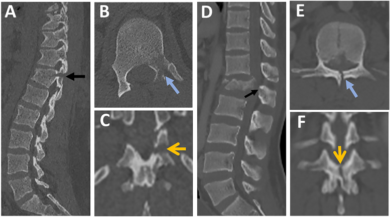
Figure 13.
The distinction between horizontal and vertical laminar fractures in CT. (A-C) images from the same patient showing (A) Parasagittal CT image showing a displaced fracture of lamina and pedicle ( black arrow); (B) Axial CT image showing a fracture of the left lamina and pedicle ( blue arrow); (C) Coronal reconstruction images showing left horizontal lamina fracture (yellow arrow); (D-F) images from the same patient showing (D) Parasagittal CT image showing a fracture of lamina and base of the spinous process (black arrow); (E) Axial CT image showing a vertical fracture of the lamina ( blue arrow); (F) Coronal reconstruction images showing the vertical orientation of the laminar fracture (yellow arrow). Abbreviations: CT, computed tomography.

