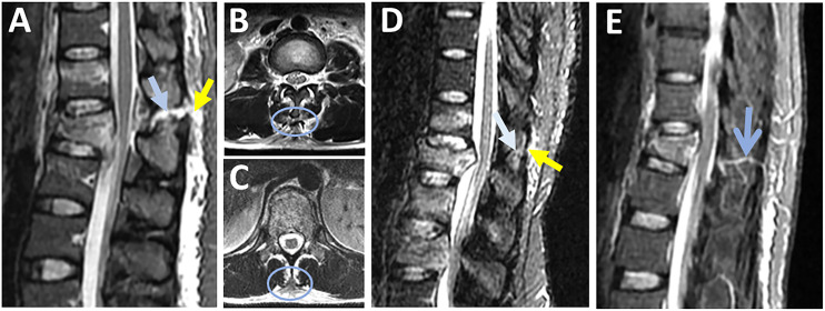
Figure 5.
Magnetic resonance imaging findings in supraspinous ligament injury. (A) Sagittal STIR images are the best sequence to detect the discontinuous black stripe due to supraspinous ligament rupture (yellow arrow) associated with high signal intensity due to interspinous ligament rupture (blue arrow); (B & C) Axial T2-weighted images show supraspinous ligament stripped from the spinous process tip (blue ring, B) or the absence of the signal intensity corresponding to the supraspinal ligament (blue ring, C ): both indicating supraspinous ligament rupture; (D) Sagittal T2-STIR image shows thinned, but continuous black stripe due to supraspinous ligament partial injury (yellow arrow) and high signal intensity due to interspinous ligament edema (white arrow); (E) vascular marking due to a crossing blood vessel (blue arrow) should not be mistaken for black stripe discontinuity. Abbreviations: STIR: short tau inversion-recovery.

