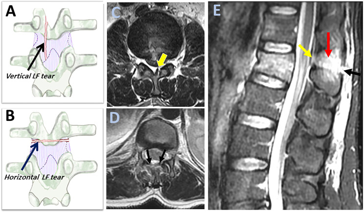
Figure 6.
Magnetic resonance imaging findings of ligamnetum flavum injury. (A-B) An illustration of the coronal view of a vertebra with the dotted lines indicates the silhouettes of the laminae and inferior articular processes behind the ligament; showing a vertical laminar fracture with an underlying vertical tear in ligamentum flavum ( black arrow, A) and a horizontal laminar fracture with an underlying horizontal tear in ligamentum flavum (blue arrow, B); (C-D) Axial T2 image can help to distinguish between horizontal and vertical ligamentum flavum it tear as it shows unilateral focal ligamentum flavum tears for vertical tears (green arrow, C) and a diffusely macerated tears for horizontal tear ( two black arrows, D); (E) Sagittal STIR image shows black stripe discontinuity due to horizontal ligament flavum tear (green arrow) and supraspinous ligament rupture (black arrow) and high signal intensity due to interspinous ligament rupture (red arrow). Horizontal ligamentum flavum tear is more easily visualized in sagittal STIR in multiple slices than the vertical ligament tear. Abbreviations: STIR: short tau inversion-recovery.

