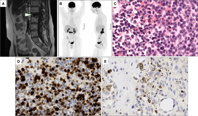
Figure 2.
Radiological and histological features of the vertebral metastases. (A) T1-weighted magnetic resonance image of the hypointense L2 spinal metastatic lesion (arrowhead). (B) 18F-fluorodeoxyglucose-positron emission tomography-computed tomography (18F-FDG-PET/CT) scan imaging revealing a hypermetabolism of the L2 spinal metastasis. (C) Microscopic examination of the pretreatment vertebral metastasis corresponding to an anaplastic melanocytic neuroectodermal transformation (MNT). Cells are pleiomorphic and numerous mitoses are visible (40×, hematoxylin and eosin [H&E] stain). (D) Ki67 immunohistochemistry in the same pretreatment vertebral biopsy showing an important proliferative index of the tumoral cells. Brown nuclear staining highlights Ki67-positive tumor cells and granular cytoplasmic brown corresponds to melanin pigments. (E) Ki67 immunohistochemistry of the post-therapy vertebral cell highlighting the presence of a residual MNT component with a lower proliferative index.

