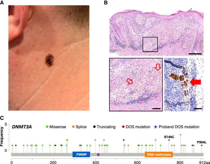
Figure 1.
Invasive melanoma in a DNMT3A overgrowth syndrome (DOS) patient. (A) Clinical appearance of a thin plaque with irregular borders and heterogeneous pigmentation on the left lateral neck. (B) Hematoxylin and eosin (H&E)-stained (top) punch biopsy specimen consisting of atypical melanocytes with at the dermal–epidermal junction, as well as pagetoid spread and an invasive dermal component, measuring 1.3 mm in thickness (scale = 250 µm) with higher magnification of the invasive component with a mitotic cell highlighted by hollow red arrows (scale = 100 µm; boxed inset and lower left panel) and MART1 stain highlighting metastatic melanoma (solid red arrow) in the sentinel lymph node (scale = 50 µm; bottom right). Sequencing was performed on the other half of this bisected specimen. (C) DNMT3A variants in melanoma curated from cBioPortal (Cerami et al. 2012; Gao et al. 2013). Proband F414fs* germline mutation is notated with a star, whereas the acquired DNMT3AR749C melanoma mutation is additionally a mutation previously described in DOS patients.

