Fig. 2.

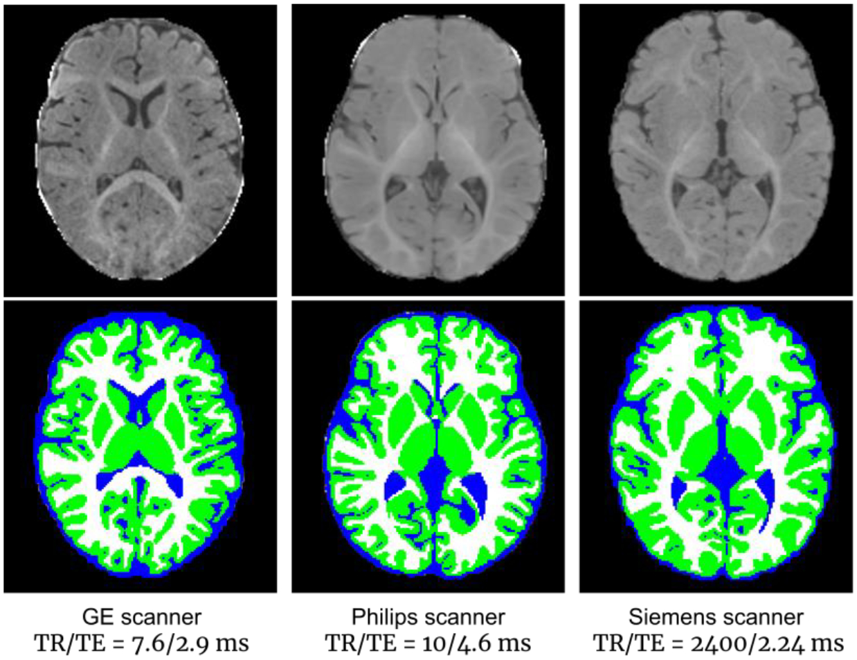
Three 6-month-old brain images acquired with different scanners and imaging protocols, showing highly variable appearance patterns (upper row), with tissue segmentation maps by iBEAT V2.0 (lower row).

Three 6-month-old brain images acquired with different scanners and imaging protocols, showing highly variable appearance patterns (upper row), with tissue segmentation maps by iBEAT V2.0 (lower row).