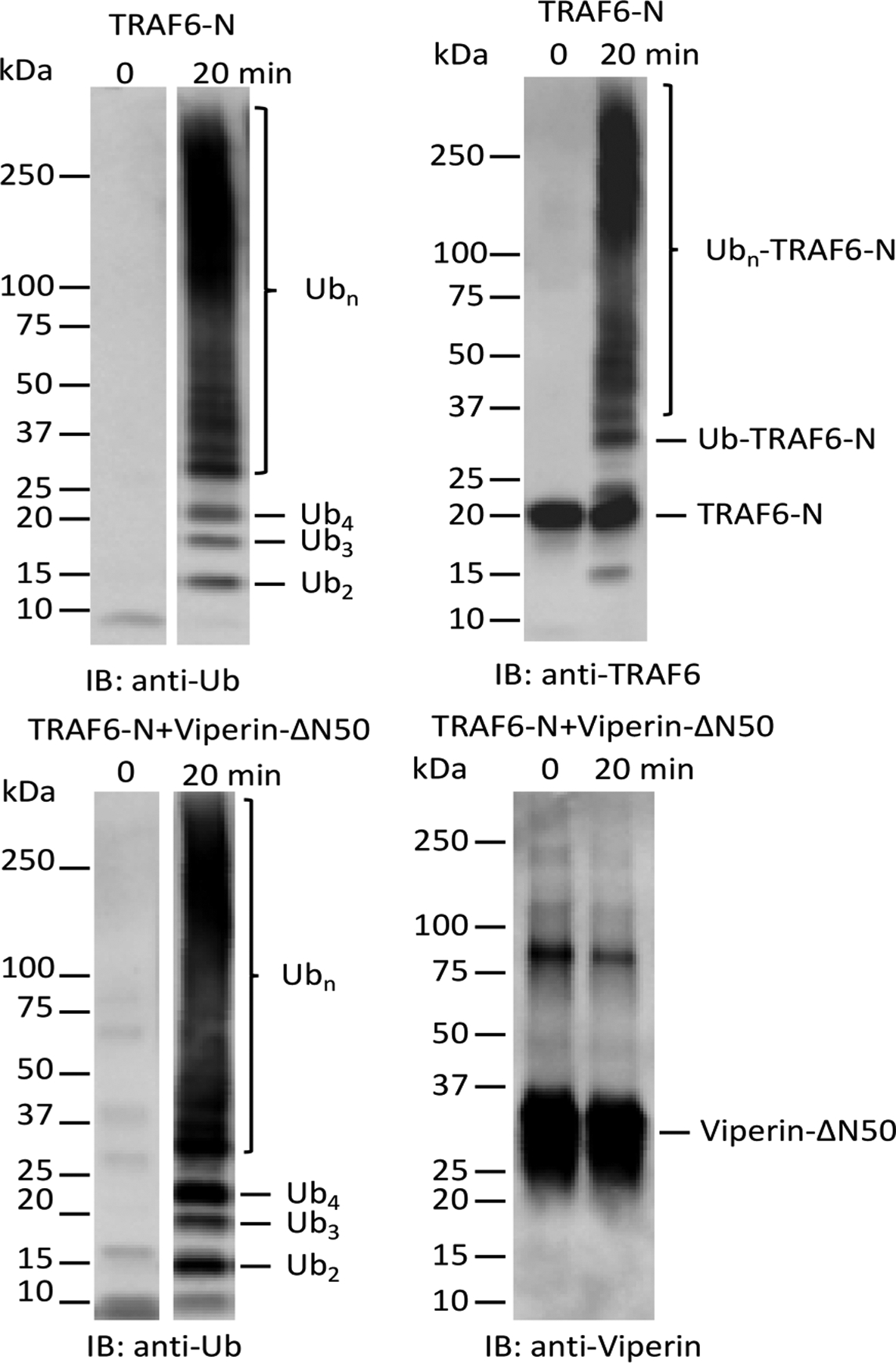
Figure 4.

Immunoblot analysis of ubiquitination reactions. Top: Staining for ubiquitin (left) and TRAF6 (right) in reactions containing TRAF6-N. Bottom: Staining for ubiquitin (left) and viperin (right) in reactions containing TRAF6-N and viperin. (Note: the polyclonal antiubiquitin antibody used in staining recognizes monoubiquitin very poorly; both t = 0 and t = 20 min lanes contain similar amounts of ubiquitin.).
