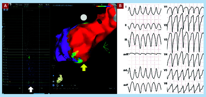
Figure 1. A. Voltage map showing a dense scar on the posterior wall of the left ventricle (yellow arrow), fragmented potentials were found in this area (white arrow). Ablation was performed with the substrate modulation technique. This study corresponds to a 49-year-old male patient with dilated ischemic cardiomyopathy. B. Clinical VT of the patient, the QRS is negative in V1 with superior axis, its origin was from the lower basal wall of the left ventricle.

