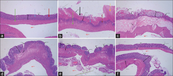
Figure 2.
Microscopic image of colon tissue after H and E staining (40 × magnification) in rats. (a) Normal tissue had normal mucosal (blue arrow) and sub-mucosal layer (green arrow), intact crypts (red arrow) with no leukocyte infiltration. (b) Control colitis group had crypt damage, mucosal layer destruction and necrosis (black arrow), thickness of sub-layer and leukocyte infiltration (yellow arrow). (c) Colitis treated with S. striata aqueous extract (SSAE, 600 mg/kg), (d) Colitis treated with S. striata hydroalcoholic extract (SSHE, 600 mg/kg), (e) Colitis treated with mesalazine (100 mg/kg) and (f) Colitis treated with dexamethasone (1 mg/kg) represent different significant degree of epithelial regeneration, diminished inflammation, leukocyte infiltration and crypt damage with no sign of necrosis

