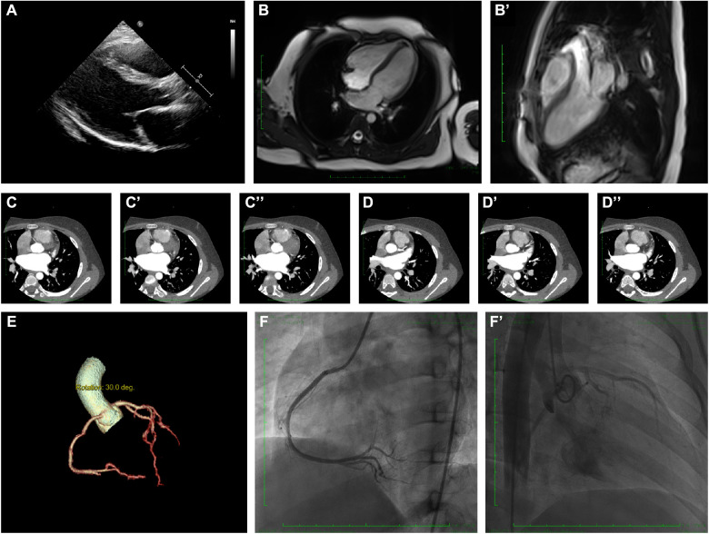
Figure 2.
Clinical and radiographic manifestation of the current proband. (A) Echocardiography revealed a slight enlargement of the left ventricle. (B–B’). Cardiac magnetic resonance showed myocardial edema in the lateral ventricular wall and apex, indicating localized injures of the myocardium. (C–C”). CTA revealed normal right coronary artery structure. (D–D”) CTA revealed normal left coronary artery structure. (E) Coronary artery rebuilding based on CTA. (F–F’) Angiographic images of the left coronary artery; no significant positive result was recorded.

