FIGURE 1.

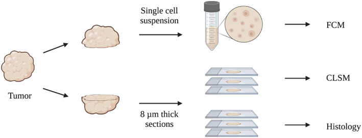
Study overview. Tumor tissue was cut in two, one part for single‐cell suspensions and subsequent analysis by flow cytometry. The other part was frozen and sectioned and imaged by confocal microscopy after staining.

Study overview. Tumor tissue was cut in two, one part for single‐cell suspensions and subsequent analysis by flow cytometry. The other part was frozen and sectioned and imaged by confocal microscopy after staining.