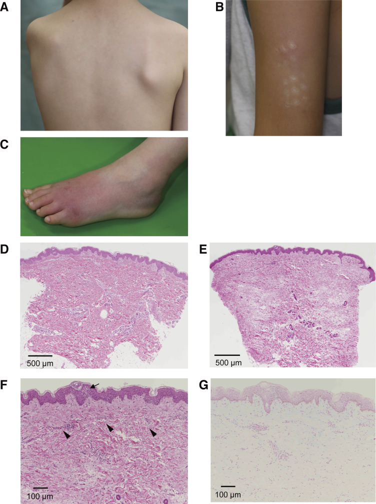
Figure S5.
Skin manifestation and pathological findings at skin biopsy of P4. (A–C) Subcutaneous nodule on the trunk (A) and rash on the left upper arm (B) and left dorsum of the foot (C). (D–G) H&E staining (D–F) and Alcian blue staining (G) of skin biopsies. The pathological findings showed slight infiltration of mononuclear cells around the small vessels of the upper dermis of the trunk (D) and in the samples from the upper dermis of the left upper arm (E; scale bar is 500 μm; original magnification, 2×); higher magnification (F) clearly showed some spongiosis in the epidermis (arrow) and slight infiltration of mononuclear cells around the small vessels (arrowheads). Sparse spaces between collagen fibers observed in F were not stained with Alcian blue (G; scale bar is 100 μm; original magnification, 40×), which may be an artifact of specimen preparation.

