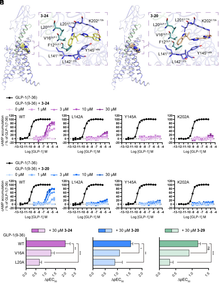
Fig. 6.
Characterization of the binding pocket of New PAMs. (A and B) Molecular docking models showing the binding mode of 3-24 (A) and 3-20 (B) in the extracellular allosteric pocket of the GLP-1R. Hydrogen bonding and cation–π interactions are indicated by dashed lines. (C and D) Potentiation of the cAMP accumulation produced by GLP-1(9-36) in combination with different fixed concentrations of 3-24 (C) or 3-20 (D) using wild-type (WT) or different mutant GLP-1R-transfected cells. (E) Comparison of the potentiation effects of specific PAMs on wide-type (WT) and mutant GLP1(9-36). ΔpEC50 represents the subtraction of pEC50 of GLP1(9-36) in the presence of a specific PAM with pEC50 of GLP1(9-36) alone. A largerΔpEC50 indicates a stronger potentiation effect of the test PAM. Data represent means ± SEM of three independent experiments performed in triplicate. Statistical analyses were performed using a two-way ANOVA test. *P < 0.05; **P < 0.01; ***P < 0.001; ns, no significant difference.

