
Figure 7.
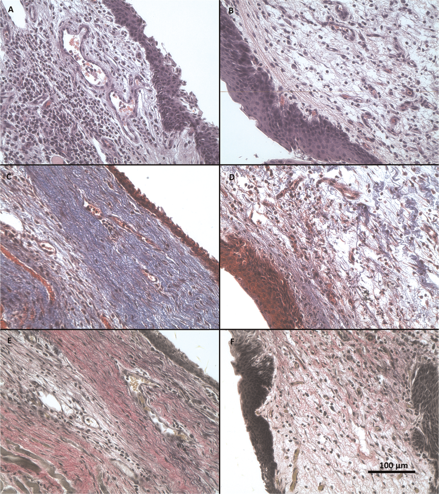
Representative vocal fold micrographs of female rabbit. Left column: operated vocal fold at the site of implantation. Right column: contralateral unoperated vocal fold. (A, B) Hematoxylin and eosin. (C, D) Masson’s trichrome. (E, F): Verhoeff’s elastic van Gieson. All at 40× magnification.