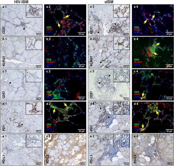
Fig. 2.
Immunohistochemistry and double immunofluorescence staining of HIV-IBM and sIBM. HIV-IBM cases showed no KLRG1+ or CD57+ cells in contrast to the sIBM cases (b1–4, c1–4). Many CD8+/CD27+ and CD3+/PD1+ cells were detectable in both groups (a1–4, d1–4). The ligands showed a weak expression for PD-L1 (e1, e3) and a non-specific staining pattern involving the endomysial connective tissue and myocyte sarcolemma cells for PD-L2 (e2, e4)

